Pacemaker Patient Education Lecture 2: Reasons for Pacemaker Implantation

This is the second in a series of short (less than 5minutes), educational videos designed for patients and their care providers to develop a thorough understanding of pacemakers.  Lecture 2 Reasons for Pacemaker Implantation describes some of the common reasons patients undergo pacemaker implantation.

Your care providers have extensive training assessing the reasons—also called indications—that a patient may need a pacemaker. In particular, it is very important that the benefits of pacemaker implantation outweigh the risks of the pacemaker implant surgery (to be discussed later). The American College of Cardiology (ACC) is one of the major professional societies that develops guidelines to help care providers make educated clinical decisions that are based upon prior clinical studies. This is the basis of “evidence-based” medicine: the process by which clinical ideas are tested, reported, and reevaluated to decide the most appropriate care for a particular condition.

The ACC has developed guidelines that help care providers decide when a patient would be best served by a pacemaker.  The easiest rule to remember is: pacemakers are most appropriate for patients who are having symptoms related to an abnormally slow—or at times, fast—heart rate. These symptoms include: shortness of breath, chest pain, dizziness, fainting (also called syncope), heart failure, arrhythmias (such as ventricular tachycardia/fibrillation), or fatigue.   The decision to implant a pacemaker also requires evaluation of the permanence of the AV block. Electrolyte abnormalities (like potassium) can cause significant AV block, but correction of the abnormality can lead to resolution of the AV block. Some diseases—like Lyme Disease—often follow a natural course where the AV block is temporary and resolves as the disease is treated. Some types of AV block that occur during periods of vagal activation can reverse very quickly (e.g., nausea and dizziness during a blood draw may cause transient AV block or during sleep in patients with sleep apnea). In addition, after aortic valve surgery, inflammation can cause transient AV block that resolves within days of the operation. Finally, there are some diseases that warrant pacemaker implantation, because the AV block may continue to worsen (for example, sarcoidosis, amyloidosis, or neuromuscular diseases).


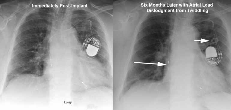
Don’t Be A Twiddler: Atrial Lead Dislodgement from Twiddler’s Syndrome

Twiddling Atrial Lead Dislodgment

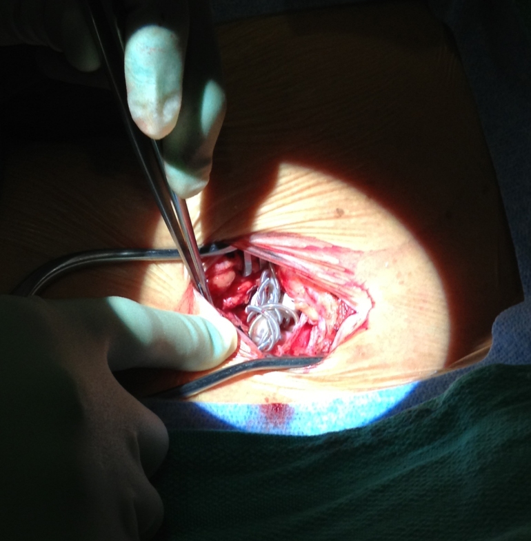
Twiddler Intraop

This elderly patient presented 6months after a dual chamber pacemaker was implanted due to symptomatic chronotropic incompetence.  They reported recurrence of exertional shortness of breath that was experienced before pacemaker implantation.  Device interrogation revealed complete loss of capture in the atrial lead. The top Figure depicts lead orientation before (left) and after (right) patient twiddling resulted in loss of slack in both leads with frank atrial lead dislodgement.  Arrows show the dislodged atrial lead and knotted leads.  The bottom Figure shows the intraoperative finding of knotted leads in the pocket.  The leads were carefully unknotted, stylets placed, and leads repositioned with normal parameters.  Originally described in 1968 [1], twiddling refers to patient manipulation of pacemaker can or leads that may lead to malfunction.  It has a reported incidence of 0.07% in a series of 17000 patients. [2]  The patient underwent an uneventful lead revision by repositioning the atrial lead and adding slack to the ventricular lead.

1  Bayliss CE, Beanlands DS, Baird RJ, “The pacemaker-twiddler’s syndrome: a new complication of implantable transvenous pacemakers,” Can Med Assoc J, V. 99 (1968), pp. 371–3.

2  T. Fahraeus and C. J. Hoijer, “Early pacemaker twiddler syndrome,” Europace, Vol. 5 (July 2003), pp. 279-281.

Pacemaker Patient Education: The Basics of Heart Anatomy and Conduction System

This is the first in a series of short (less than 5minutes), educational videos designed for patients and their care providers to develop a thorough understanding of pacemakers.  Lecture 1 The Basics of Heart Anatomy and Conduction System introduces the core concepts of heart function necessary to understand the role of pacemakers in the management of heart disease.

Please check back for future lectures to be posted and register to receive updates from the Heart Rhythm Center.

Lecture 2: Reasons for Pacemaker Implantation

Lecture 3: What are Pacemakers and How Do They Work?

Lecture 4: Preoperative Workup and Evaluation

Lecture 5: Meeting the Implanting Physician

Lecture 6: The Implant Procedure

Lecture 7: Possible Complications of Pacemaker Implantation

Lecture 8: Post-operative Care of the Pacemaker Patient (The First Month)

Lecture 9:  Long-Term Care and Follow-up of the Pacemaker Patient

Lecture 10:  What are Pacemaker Device Recalls/Advisories/Alerts?

New Book for Patients that Need or Have Undergone Pacemaker Therapy

I wrote What is a Pacemaker? Cardiologist’s Guide for Patients and Their Care Providers to fill a gap in available resources for patients who have undergone or are under evaluation for pacemaker implantation. Please consider this a resource for your patients, colleagues, friends, and family.

The fastest growing population segment in the United   States, seniors commonly undergo pacemaker implantation. Although doctors’ offices typically provide short pamphlets on pacemaker implantation, there is rarely any comprehensive yet understandable reference material for the patients to obtain…until now. Explaining the “what, why, and how” of pacemaker implantation, this invaluable new guide provides an in-depth summary of pacemakers, from the initial patient evaluation and device implantation to the issues that could potentially arise during a long-term follow up.

Feasibility Study for Leadless Pacemaker Presented at Heart Rhythm Society

A study revealed at this year’s Heart Rhythm Society Meeting presented the first in-human results of a leadless implantable pacemaker. The device is about the size of a AAA battery and is implanted in the right ventricle. A limitation of current pacemakers is the reliance on implantable leads that can fracture or become infected. This device is the first step toward developing leadless pacing technologies. It remains to be seen how clinically useful this device will be but is expected to be available in Europe later this year.

Complications of Pacemaker Implantation

Approximately 180000 patients undergo pacemaker implantation in the U.S each year [1]. In addition, the extreme elderly are the most rapidly growing segment of the U.S. [2,3] and pacemakers are commonly implanted in this population. There are reports of pacemaker implant complications (generally clinical trials reporting outcomes and incident complication rates) and fewer reports of complication rates in the extreme elderly (with a persistent exclusion of elderly patients from ongoing clinical trials [4]). A comprehensive review of pacemaker implant complications can help improve informed consent in preoperative patients. Major and minor complications are defined based upon prior reports of device-related complications. [5,6,7,8] Major complications have been defined as death, cardiac arrest, cardiac perforation, cardiac valve injury, coronary venous dissection, hemothorax, pneumothorax, transient ischemic attack, stroke, myocardial infarction, pericardial tamponade, and arterial-venous fistula. Minor complications have been defined as drug reaction, conduction block, hematoma or lead dislodgement requiring reoperation, peripheral embolus, phlebitis, peripheral nerve injury, and device-related infection. This chapter will include discussion of common and uncommon complications of pacemaker implantation including associated incidence as well as the associated radiographs and common clinical signs of these complications.
Complications of Pacemaker Implantation

How are Pacemaker Leads Implanted?

The heart’s natural pacemaker (the SA node) is located in the top right chamber of the heart, the right atrium. The SA node sends a signal to the upper right (right atrium) and left chambers (left atrium) and lower chambers (the right and left ventricles) via the atrioventricular (AV) node. Often, the patient has a slow heart rate because the electrical connection between the top (the signal from the SA node) and bottom (the ventricles) of the heart (right ventricle) is diseased and a pacemaker lead can be placed in this chamber.

The most common type of pacemaker involves placing a lead in the right atrium and right ventricle.  The image above (taken from MedtronicConnect.com) displays a pacing lead in the right atrium and right ventricle.  It also shows a typical implant scenario where the device is positioned under the left clavicle below the skin but above the chest muscle.  The leads are then placed through the subclavian vein where they are threaded to position into the heart.  The following video (taken from MedtronicConnect.com) shows the sequence of events during lead implantation.

You can see the site of the incision is under the left collarbone and insertion of the needle, guide wire, and introducer (hollow tube used to carry lead into the vein).  The pacing lead is then advanced into the right ventricle where the screw-in mechanism is activated – views of both the helix extending out and the implant tool operation are shown.

Difficult Access of Coronary Sinus During Attempted Percutaneous LV Lead Insertion

The emergence of resynchronization therapy has led to an increase in attempts at left ventricular lead placement via the coronary sinus (CS).  The MIRACLE study program [LEO05] reported a 91.6% success rate for LV lead placement, while COMPANION [BRI04] revealed an 89%  success rate for LV lead placement.  Another report indicated a similar 92% success rate with LV lead placement. [DIV08]  Though we counsel our patients on a LV lead placement success rate at 88-92%, our center demonstrated a 97% success rate (64 of 66 patients) with LV lead placement within the range from 2:30 to 5:30 o’clock in the left anterior oblique (LAO) view. [WIL10]

There are many reasons for difficult CS access including Thebesian valves, cardiac vein ostial valves, and Chiari networks.  The image shown below is representative of the ostial valve of the coronary sinus (aka, Thebesian valve) that prevents engagement of the CS and ultimately precludes LV lead placement.  Routine attempts at engaging the coronary sinus with deflectable EP catheters as well as hydrophilic guide wires were met without success.  A 9MHz radial intracardiac echo (ICE) probe (UltraICE, Boston Scientific Corp) was introduced via a steerable sheath (Agilis, St. Jude Medical) to evaluate the CS anatomy.  This image demonstrates a Thebesian valve covering the entire CS osmium with no obvious accessible fenestration or defect.  LV lead placement was aborted and patient was referred for epicardial LV lead placement via cardiothoracic surgery.


Prior reviews of CS anatomy (PEJ08) revealed the presence of Thebesian valves in 80% of cases and Chiari networks were found in 10% of cases.  It covered one-fifth in 7%, one-third the os in 29%, one-half in 27%, two-thirds in 14%, and the entire os in 5%.  The average diameter of the CS os was 8mm with a range of 3-15mm.  Appreciation for the anatomic variations of the normal human CS as well as experience with ICE may help reduce complications and improve success of LV lead implantation.

References:

LEO05     Leon AR, Abraham WT, Curtis AB, et al.; for the MIRACLE Study Program, “Safety of Transvenous Cardiac Resynchronization System Implantation in Patients with Chronic Heart Failure: Combined Results of Over 2000 Patients from a Multicenter Study Program,” J Am Coll Cardiol, 2005;46(12):2348–56.

BRI04     Bristow MR, Saxon LA, Boehmer J, et al., “Cardiac-Resynchronization Therapy with or without an Implantable Defibrillator in Advanced Chronic Heart Failure for the Comparison of Medical Therapy, Pacing, and Defibrillation in Heart Failure (COMPANION) Investigators,” N Engl J Med, 2004;350(21):2140–50.

DIV08     D’Ivernois C, Lesage J, Blanc P, “Where are left ventricular leads really implanted? A study of 90 consecutive patients,” Pacing Clin Electrophysiol, 2008;31(5):554–9.

WIL10     Williams JL, Lugg D, Gray R, Hollis D, Stoner M, Stevenson R, “Patient Demographics, Complications, and Hospital Utilization in 250 Consecutive Device Implants of a New Community Hospital Electrophysiology Program,” American Heart Hospital Journal, V. 8, No. 1 (Summer, 2010), pp. 33-39.

PEJ08     Pejkovic B, Krajnc I, Anderhuber F, Kosutic D, “Anatomical Variations of the Coronary Sinus Ostium Area of the Human Heart,” J Int Med Research, V. 36 (2008), pp. 314-321.

Heart Rhythm Management and Drug Delivery via Implantable Biosensors

Your Next Prescription Might Be For A Microchip – Forbes.

Drug delivery via implantable biosensors is the next generation of devices that can be used to help heart rhythm disorders.  Our current implantable loop recorders allow us to track heart rhythm disorders and link symptoms to arrhythmias.  Implantable biosensors will combine this monitoring ability with automated drug delivery.

Pacemaker Implantation in the Extreme Elderly

Right Atrial (A), Right Ventricular (B), and Left Ventricular (C) Leads Before (Pre) and After (Post) Dislodgements. Right atrial lead became dislodged after patient twiddled with device. Right ventricular lead dislodged by moving more basilar in position (arrow) one day after implant. Left ventricular lead dislodged and reseated itself in the body of coronary sinus 3 months after initial placement (arrow).

     There are scant data for pacemaker implant complications and readmission rates in the extreme elderly (age≥80 years) despite their common use in this population.   We performed a retrospective chart review of consecutive patients (n=149, age≥80 years) who underwent pacemaker implantation at our community hospital Electrophysiology program from July 2008 through June 2010.  Single-, dual-, and biventricular-chamber pacemakers and generator changes were included for analysis; cardioverter-defibrillator devices, temporary pacemakers, and loop recorders were excluded.  Standard procedures for implantation were used.   Major complications defined as death, cardiac arrest, cardiac perforation, cardiac valve injury, coronary venous dissection, hemothorax, pneumothorax, transient ischemic attack, stroke, myocardial infarction, pericardial tamponade, and arterial-venous fistula.  Minor complications defined as drug reaction, conduction block, hematoma or lead dislodgement requiring reoperation, peripheral embolus, phlebitis, peripheral nerve injury, and device-related infection.

     The overall mean age of implantation was 86 years.  There were no intraprocedural complications. There was one major in-hospital (0.7%) and one minor in-hospital complication (0.7%).  Within 30 days of implant, there was an overall 5.4% rate of complications; 4 minor (2.7%) and 4 major (2.7%).  There was a 30d cardiovascular-attributable mortality of 0.7% and an all-cause mortality of 2%.  There was a 5.4% rate of readmission within 30days of implantation.

Our report of pacemaker implantations in the extreme elderly reveals rates of implant complications comparable to data from younger patient populations while experiencing a higher 30day all-cause mortality (that may be attributable to elevated all-cause mortality rates in this age-group).