Saving money and improving quality should be a priority no matter how “small” the impact. By 2028, Medicare Trustees project Medicare does not meet either the Trustees’ short-range test of financial adequacy or long-range test of close actuarial balance.

Yiyan Liu and I presented our research today at ACC 2023 Annual Sessions.

Background: Prior modeling has demonstrated the extended battery longevity of biventricular defibrillator (CRT-D) reduced additional implant or replacement procedures, yet real-world evidence on health economics data is limited. This study reports the total direct medical costs to Medicare associated with CRT-D procedures.

Methods: A retrospective study was conducted using the 5% Medicare Standard Analytical Files (SAF). Patients with CRT-D implant or replacement between 1/2009 and 12/2020 in hospital settings were identified using ICD-9-PCS (00.54) and ICD-10-PCS (0JH609Z, 0JH639Z, 0JH809Z, 0JH839Z) codes supplemented with Current Procedural Terminology* (CPT®) codes (33225, 33249, 33263, 33264). Total direct medical costs to Medicare included costs for all claim types. To capture medical costs leading to and following the procedure, the costs were calculated for three time periods: a 30-day interval before the procedure, the procedure encounter period, and a 30-day interval after the procedure. All analyses were performed using the Instant Health Data (IHD) software (Panalgo, Boston MA, USA) and R, version 3.2.1 (R Foundation for Statistical Computing, Vienna, Austria).

Results: Among the 15,002 Medicare patients who underwent CRT-D implant or replacement from 2009 through 2020, the mean age at first procedure was 72 and 71% were male. The number of patients with 1, 2, and 3 CRT-D procedures was 12,944, 1,923, and 135, respectively. The total cumulative cost to Medicare for an average patient undergoing 1, 2, and 3 generator implant or replacement procedures was $52,795, $88,976, and $128,846 in 2021 dollars, respectively (Figure).

Discussion: Battery capacity as measured in ampere-hours (Ah) is the strongest predictor of CRT-D battery longevity. Extended defibrillator battery longevity is preferred by patients and cost savings for health care budgets. Data have demonstrated extended CRT-D extended battery life exceeded patient survival in a typical HFrEF cohort.1 These extended longevity CRT-D devices not only outlast patient life expectancy but avoid costs of complications and generator changes. The data presented here demonstrate the potential for a significant cost savings when fewer generator changes are required in Medicare patients due to battery depletion and when clinically appropriate.

CRT-D replacement due to battery depletion is a significant cost-driver for payors2,3 and a significant complication-driver for patients.4,5 Landolina et al. found the need for device replacements at six years was reduced from 83% to 68% with the use of devices with improved battery longevity from the most recent generation.2 Modeling has shown increased utilization of extended longevity CRT-D led to a 39% annual reduction in major complications (n=1099) and a 12.8% reduction in total annual costs ($496million) for Medicare.6 The data presented here indicate a 244% increase in cost when three CRT-D generator implant/replacement procedures were completed versus one were performed among 15,002 Medicare patients who underwent CRT-D implant or replacement from 2009 through 2020.

Limitations: This research used the number of generator implants/replacements as a surrogate for using extended battery longevity CRT-D; certainly, there will be incidences of device infection rather than battery depletion as indication for generator replacement. Prior data has shown the battery depletion is the most common cause of generator replacement even when using extended longevity CRT-D.6 These costs do not account for the nonfinancial or clinical outcome of additional complications resulting from more frequent generator changes. Finally, it is difficult to quantify the underlying systemic conflicts of interest where frequent CRT-D generator changes continue to drive fee-for-service or productivity-based reimbursements for physicians and health systems.

Conclusion: The total direct medical costs to Medicare for CRT-D implant or replacement increased substantially with increased procedure frequency.

References:

  1. Williams JL, Harley B, Williams G, “First Demonstration of Cardiac Resynchronization Therapy Defibrillator Service Life Exceeding Patient Survival in a Heart Failure with Reduced Ejection Fraction Cohort,” J Innov Cardiac Rhythm Manage. 2020; 11(12): 4325–4332.
  2. Landolina M, Morani G, Curnis A, et al. The economic impact of battery longevity in implantable cardioverter-defibrillators for cardiac resynchronization therapy: the hospital and healthcare system perspectives. Europace. 2017;19(8):1349–1356.
  3. Gadler F, Ding Y, Verin N, et al. Economic impact of longer battery life of cardiac resynchronization therapy defibrillators in Sweden. Clinicoecon Outcomes Res. 2016;8:657–666.
  4. Poole JE, Gleva MJ, Mela T, et al. Complication rates associated with pacemaker or implantable cardioverter-defibrillator generator replacements and upgrade procedures results from the REPLACE registry. Circulation. 2010;122(16):1553–1561.
  5. Prutkin JM, Reynolds MR, Bao H, et al. Rates of and factors associated with infection in 200,909 medicare implantable cardioverter-defibrillator implants: results from the NCDR®. Circulation. 2014;130(13):1037–1043.
  6. Williams JL and Williams GM, “Modeling long-term effect of biventricular defibrillator battery capacity on major complications and costs associated with replacement procedures.” Heart Rhythm Journal, V. 18, Issue 8, August 01, 2021: S396-S397.

Jeffrey Williams, MD, MS, FACC, FHRS, CPE and Gabriella Williams
James A Haley VA Medical Center and University of Notre Dame

Problem Statement or Scientific Question: There is consensus that the subcutaneous (SC) implantable cardioverter defibrillator (ICD) is associated with fewer lead-related complications albeit limited battery longevity (based on Ah) compared to transvenous (TV) ICD. The type of device implanted is a shared decision made by the implanting physician and patient however, the long term costs of the SC versus TV ICD when balancing complications and battery longevity are unclear.

Background/Project Intent: To model the cost and complication rate variations between the 0.8Ah SC ICD and 1.0Ah, 1.9Ah, and 2.0Ah battery single chamber TV ICD generator replacements in a model over 26 years.

Methodology: A model was developed using MATLAB (Mathworks Inc., Natick, MA) estimating the rate of generator replacements of the SC ICD versus the TV-ICD (using 20% utilization for each device) to assess differences in costs and complications over time. Battery longevities were based upon prior “real-world” studies of CRT-D and extrapolated to single chamber TV ICD based upon current literature. Costs and baseline volumes for primary prevention ICD in the United States were based upon publicly available estimates. Model parameters including heart failure prevalence, mortality, and complication rates were based upon prior data.

Results: The 2.0Ah TV ICD had fewer generator replacements (max n=7200 annually) and costs (max annual savings of $247,500,000) compared to the SC ICD, 1.0Ah, and 1.9Ah TV ICD. The SC ICD had less generator replacement costs compared to the 1.0Ah and 1.9Ah TV ICD. The SC ICD was associated with fewer annual complications (max n=422) compared to all TV ICD.

Value Proposition: The 2.0Ah TV ICD reduced the total number of implant procedures for the patient. The 2.0Ah TV ICD was associated with lower costs for payers and society. The SC ICD had the fewest long-term complications.

Conclusions: The 2.0Ah TV ICD is associated with fewer replacement procedures and costs than smaller battery capacity TV ICD as well as SC ICD. The SC ICD had the fewest long-term complications when compared to the TV ICD (all battery capacities). The decision on device implant type requires a complex informed consent discussion with the patient to address costs, number of replacement procedures, and overall long-term complication rates.

References:

Alam MB, Munir MB, Rattan R, Adelstein E, Jain S, Saba S. Battery longevity from cardiac resynchronization therapy defibrillators: differences between manufacturers and discrepancies with published product performance reports. Europace. 2017;19(3):421–424.

Williams JL and Williams GM, “Modeling long-term effect of biventricular defibrillator battery capacity on major complications and costs associated with replacement procedures.” Heart Rhythm Journal, V. 18, Issue 8, August 01, 2021: S396-S397.

Poli S, Boriani G, Zecchin M, FacchinD, Gasparini M, Landolina M, Pietro Ricci R, Lanera C, Gregori D, and Proclemer A, “Favorable Trend of Implantable Cardioverter-Defibrillator Service Life in a Large Single-Nation Population: Insights From 10-Year Analysis of the Italien Implantable Cardioverter-Defibrillator Registry. J Am Heart Assoc. 2019;8:e012759.

Cutler D et al, “Physician Beliefs and Patient Preferences: A New Look at Regional Variation in Health Care Spending,” Amer Econ Jour: Econ Policy, Vol 11, No 1, Feb 2019, pp. 192-221.

Caverly TJ et al, “Patient Preference in the Decision to Place Implantable Cardioverter-Defibrillators,” Arch Intern Med. 2012;172(14):1104-1107.

Please watch my short presentation given at the 2021 Annual Sessions of the Heart Rhythm Society on July 30, 2021. A 2.1 ampere-hour (Ah) battery capacity biventricular defibrillator (CRT-D) was recently shown to be the first to exceed patient survival in a chronic heart failure cohort. There is consensus that extended battery longevity (based on Ah) can reduce the costs and complications related to generator replacements. Our model revealed that increased utilization of extended longevity CRT-D battery technology (2.1Ah) leads to substantial annual reductions in major complications and costs of replacement procedures.

We developed a model to estimate cost and complication rate variations associated between 1.0Ah, 1.6Ah, and 2.1Ah battery CRT-D generator replacements over a period of 20 years.

Introduction: From the initial report of intraoperative radiofrequency (RF) ablation causing esophageal injury,GIL01 atrioesophageal fistulas (AEF) have been reported in percutaneous atrial fibrillation RF ablations.SCA04,PAP04 Atrioesophageal fistulas have been estimated to occur in as many as 1% of AF ablationsDOL03 but a likely accepted event rate is less than 0.1%.PAP04,SCA04,CUM06 The mortality associated with AEF is devastating and was found to approach 100% in the largest published registry of AEF.CUM06 This is in stark contrast to a near zero death rate of atrial perforations during RF ablation.BUN05 An article by Müller et al (http://www.heartrhythmjournal.com/article/S1547-5271(15)00418-X/abstract) examined the high incidence of esophageal lesions after atrial fibrillation ablations related to the use of esophageal temperature probes. Multivariate analysis revealed the use of the temperature probe was the only independent predictor of esophageal lesions. Finally, data in Heart Rhythm examined the rate of atrioesophageal fistula formation with contact force (CF) sensing catheters versus non-CF-sensing catheters. Black-Maier et al found the “occurrence of atrioesophageal fistula formation accounted for a 5-fold higher proportion of all MAUDE medical device reports of injury or death with CF-sensing catheters compared with non-CF-sensing catheters.”

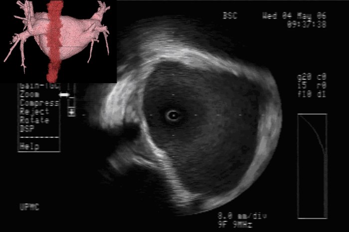
Value of Imaging: The value of intracardiac imaging via radial intracardiac echo (ICE) cannot be underestimated given the nonuniform thickness and variable course along the posterior wall of the left atrium.SAN05,GOO05,REN06 The proximity of the esophagus to the left pulmonary venous antrum is depicted in Figure 1. Typically, patients with AEF present a mean of 12.3days after their procedure;CUM06 however, presentation within 3-5days of the ablation has been reported.PAP04 Findings on CT scans can be non-specific but, infected pleural and pericardial effusions may suggest esophageal contamination of the pleural spaces. CT scan of the chest (without oral contrast) with the presence of intravenous contrast seen in the esophagus or surrounding posterior mediastinum would imply a fistulous connection.MAL07 Additionally, one may note a narrowed, irregular, and ulcerated pulmonary vein, posterior left atrial wall thickening, posterior mediastinal fat induration, or pneumomediastinum.MAL07

Imaging Demonstrated Proximity of Esophagus to Left Pulmonary Veins. The top left inset of the figure depicts the 3D reconstruction of the left atrium and pulmonary veins with the esophagus tagged in red. The course of the esophagus is along the posterior left atrium in contiguity to the left pulmonary vestibule. The ICE image of the left pulmonary vestibule shows the characteristic echocardiographic signature of an esophageal temperature probe in the 8 o’clock position.

Mechanism of Esophageal Injury: Finite-element analysis supports that esophageal injury is exclusively due to thermal conduction from the atrium.BER05 Esophageal injury can occur despite small electrode size, low power (<30W), and low electrode temperature (34°C). There are two caveats however, irrigated electrodes and electrode-endocardial contact verification (direct visualization with ICE or force-sensing) may increase power delivery to the tissue.

Avoiding Esophageal Injury: There has been much enthusiasm to determine means by which esophageal injury can be avoided. These include echocardiographic monitoring for microbubble formation,CUM05 the use of cryoablation to lower esophageal ulceration,RIP07 plan ablations to avoid esophagus by creating virtual esophageal tube using electroanatomic mapping,SHE07 esophageal irrigation to lower esophageal temperature,TSU07 and physically deflecting the esophagus away from the ablation site.HER06 The study by Muller et alMUL15 suggests the possibility that esophageal temperature probes may increase susceptibility to esophageal lesions. Figure 2 shows ICE images before and after orogastric tube removal. One notes the signature of the OGT at 8 o’clock in the left image. There is a small indentation in the posterior left atrial wall at the site of the OGT. On the right, imaging demonstrates this indentation is resolved after removal of the OGT. I will often remove esophageal instrumentation to avoid any possible displacement of the esophagus towards the left atrium.

Intracardiac Echocardiography (ICE) of Orogastric Tube (OGT). The left image depicts the ICE signature of the OGT at ~8 o’clock. There is a small indentation in the posterior left atrial wall at the site of the OGT. On the right, ICE demonstrates the resolution of this indentation after removal of the OGT.

CF-sensing catheters have certainly enhanced ability to deliver more consistent lesions however, there are clearly limitations when the operator cannot see real-time electrode-endocardial contact. There have been many times where I have seen left and right atrial tenting due to catheter contact at less than 10g of force. Force sensing has certainly added to our armamentarium but I would caution all that there is more to ablation than contact force.

Note:  These radial ICE images would not be possible without my mentor, Dr. David Schwartzman (Pittsburgh, PA).

REFERENCES:

GIL01  Gillinov AM, Pettersson G, Rice TW, “Esophageal injury during radiofrequency ablation for atrial fibrillation,” J Thor Card Surg, V. 122, No. 6 (December 2001), pp. 1239-1240.

SCA04  Scanavacca MI, D’Avila A, Parga J, Sosa E, “Left Atrial-Esophageal Fistula Following Radiofrequency Catheter Ablation of Atrial Fibrillation,” J Cardiovasc Electrophysiol, V. 15, No. 8 (August 2004), pp. 960-962.

PAP04  Pappone C, Oral H, Santinella V, Vicedomini G, Lang CC, Manguso F, Torracca L, Benussi S, Alfieri O, Hong R, Lau W, Hirata K, Shikuma N, Hall B, Morady F, “Atrio-Esophageal Fistula as a Complication of Percutaneous Transcatheter Ablation of Atrial Fibrillation,” Circulation, V. 109 (June 8, 2004), pp. 2724-2726.

DOL03  Doll N, Borger MA, Fabricius A, Stephan S, Gummert J, Mohr FW, Hauss J, Kottkamp H, Hindricks G, “Esophageal perforation during left atrial radiofrequency ablation: Is the risk too high?” J Thor Cardiovasc Surg, V. 125, No. 4 (April 2003), pp. 836-842.

CUM06  Cummings JE, Schweikert RA, Saliba WI, Burkhardt D, Kilikaslan F, Saad E, Natale A, “Brief Communication: Atrial-Esophageal Fistulas after Radiofrequency Ablation,” Ann Int Med, V. 144, No. 8 (18 April 2006), pp. 572-574.

BUN05  Bunch TJ, Asirvatham SJ, Friedman PA, Monahan KH, Munger TM, Rea RF, Sinak LJ, Packer DL, “Outcomes After Cardiac Perforation During Radiofrequency Ablation of the Atrium,” J Cardiovasc Electrophysiol, V. 16, No. 11 (November 2005), pp. 1172-1179.

SCH06  Schwartzman DS, Nosbisch J, and Housel Debra, “Echocardiographically guided left atrial ablation: Characterization of a new technique,” Heart Rhythm, V. 3, No. 8 (August 2006), pp. 930 –938.

SAN05  Sanchez-Quintana D, Cabrera JA, Climent V, Farre J, de Mendonca MC, Ho SY, “Anatomic Relations Between the Esophagus and Left Atrium and Relevance for Ablation of Atrial Fibrillation,” Circulation, V. 112 (September 6, 2005), pp. 1400-1405.

GOO05  Good E, Oral H, Lemola K, Han J, Tamirisa K, Igic P, Elmouchi D, Tschopp D, Reich S, Chugh A, Bogun F, Pelosi F Jr, Morady F, “Movement of the Esophagus During Left Atrial Catheter Ablation for Atrial Fibrillation,” JACC, V. 46, No. 11 (December 6, 2005), pp. 2107-21190.

REN06  Ren J-F, Lin D, Marchlinski FE, Callans DJ, Patel V, “Esophageal imaging and strategies for avoiding injury during left atrial ablation for atrial fibrillation,” Heart Rhythm, V. 3, No. 10 (October 2006), pp. 1156-1161.

MAL07  Malamis AP, Kirshenbaum KJ, and Nadimpalli S, “CT Radiographic Findings: Atrio-esophageal Fistula After Transcatheter Percutaneous Ablation of Atrial Fibrillation,” J Thorac Imaging, V. 22, No. 2 (May 2007), pp. 188-191.

BER05  Berjano EJ and Hornero F, “What affects esophageal injury during radiofrequency ablation of the left atrium? An engineering study based on finite-element analysis,” Physiol Meas, V. 26 (2005), pp. 837-848.

CUM05  Cummings JE, Schweikert RA, Saliba WI, Burkhardt JD, Brachmann J, Gunther J, Schibgilla V, Verma A, Dery MA, Drago JL, Kilicaslan F, Natale A, “Assessment of Temperature, Proximity, and Course of the Esophagus During Radiofrequency Ablation Within the Left Atrium,” Circulation, V. 112 (July 26, 2005), pp. 459-464.

RIP07  Ripley KL, Gage AA, Olsen DB, Van Vleet JF, Lau C-P, Tse H-F, “Time Course of Esophageal Lesions After Catheter Ablation with Cryothermal and Radiofrequency Ablation: Implication for Atrio-Esophageal Fistula Formation After Catheter Ablation for Atrial Fibrillation,” J Cardiovasc Electrophysiol, V. 18, No. 6 (June 2006), pp. 642-646.

SHE07  Sherzer AI, Feigenblum DY, Kulkarni S, Pina JW, Casey JL, Salka KA, Simons GR, “Continuous Nonfluoroscopic Localization of the Esophagus During Radiofrequency Catheter Ablation of Atrial Fibrillation,” J Cardiovasc Electrophysiol, V. 18, No. 2 (February 2007), pp. 157-160.

TSU07  Tsuchiya T, Ashikaga K, Nakagawa S, Hayashida K, Kugimiya H, “Atrial Fibrillation Ablation with Esophageal Cooling with a Cooled Water-Irrigated Intraesophageal Balloon: A Pilot Study,” J Cardiovasc Electrophysiol, V. 18, No. 2 (February 2007), pp. 145-150.

HER06  Herweg B, Johnson N, Postler G, Curtis AB, Barold SS, Ilercil A, “Mechanical Esophageal Deflection During Ablation of Atrial Fibrillation,” PACE, V. 29 (September 2006), pp. 957-961.

MUL15  Müller P, Dietrich J-W, Halbfass P, Abouarab A, Fochler F, Szöllösi A, Nentwich K, Roos M, Krug J, Schade A, Mügge A, Deneke T, “Higher incidence of esophageal lesions after AF ablation related to the use of esophageal temperature probes,” Heart Rhythm, Published Online: April 03, 2015.

Starting 2021 with new paper! Looking at longevity of 2.1Ah biventricular defibrillators (CRT-D) and perhaps help explain high rate of complications when patients need to undergo generator changes for battery depletion. More research is needed to examine the clinical and cost effectiveness of avoiding generator changes during a vulnerable physiologic time in the lives of CRT-D patients.

Key Points:

  • These data demonstrated the first reversal in ICD battery longevity versus patient survival; the 2.1-Ah ICD battery life exceeded patient survival in a typical HFrEF cohort.
  • Our results support the hypothesis that the acceleration of device OOS during the sixth to ninth years (when it is expected that roughly 98% of 1.0-Ah and 1.4-Ah CRT-D systems reach ERI) may explain the historically high rate of complications for ICD generator changes as compared with at the initial implantation.
  • During the entire study, only 5.7% of 2.1Ah devices reached the ERI point (average time to ERI: 7.8 ±1.5 years) in up to 10.3 years of follow-up.

Read full manuscript at Journal of Innovations in Cardiac Rhythm Management.

Welcome to the 4th Annual Lakeland Regional Health Cardiovascular Symposium. Tired of CV Symposiums that focus on the procedures that can be done to your patients rather than prevention of CV disease?!? This year we’ll be focusing on the PREVENTION of CV disease. Please click to attend the Symposium for 5.5 hours of free CME on Saturday February 8, 2020. I’ll be adding links to any talks the speaker has permitted so you can follow along on day of Symposium.

7:30 – 7:55A Registration and Continental Breakfast
7:55 – 8:00A Welcome Remarks
8:00 – 8:40A: Lipid management and risk panels for cardiovascular disease, Dr. Stephen Kopecky


8:40 – 9:30A: Prevention of Sudden Cardiac Death, Williams


9:30 – 10:00A: Prevention of Cardioembolic Stroke, Dr. Khanna


10:00 – 10:20A: Ask-the-Experts Refreshment Break
10:20 – 11:00A: DM2 and CV Disease, Dr. Owen

11:00 – 11:40A: Frequent Touch Primary Care and CV Disease Prevention, Drs. Ghany and Syed

11:40A – 12:20P: Applying Evidence-Based Guidelines to Lower Heart Failure Readmissions, Dr. Navin Rajagopalan


12:20 – 12:30P: Ask-the-Experts Refreshment Break
12:30 – 1:30P: Luncheon, Plant-Based Diets and Prevention of CV Disease, Dr. Monica Aggarwal

Many thanks to the faculty of the 2019 LRH Cardiovascular Symposium! We had over 200 registrants for 6 hours of great cardiovascular CME. From left to right, Dr. Parag Patel (Mayo Clinic), Dr. Anuja Dokras (University of Pennsylvania), Dr. Jeff Williams (LRH), Dr. Carl Pepine (University of Florida), Dr. Edward Tadajweski (WellSpan Health), Dr. Philip Owen (LRH), Dr. Matthew Martinez (Lehigh Valley Health Network), and Dr. Kathryn Lindley (Washington University). Dr. Denise Edwards (University of South Florida) is not pictured.

We had over 200 registrants to this year’s CV Symposium with physicians and nurses traveling from all over Florida. Our faculty was fantastic and their lectures are included below.

Anuja Dokras, MD, PhD, Associate Professor, Penn Fertility Care, University of Pennsylvania Medical Center, Philadelphia, PA. Dr. Dokras lectured about the role of obstetric and gynecologic issues and the future risk of heart disease in women.

Denise Edwards, MD, Director, Healthy Weight Clinic, Assistant Professor of Internal Medicine and Pediatrics, USF Health. Dr. Edwards lectured about the assessment and treatment of obesity in adolescents and women.

Kathryn J. Lindley, MD, Assistant Professor of Medicine, Director, Center for Woman’s Heart Disease, Washington University School of Medicine. Dr. Lindley spoke about the risks of women’s heart disease in pregnancy.

Matthew W Martinez, MD FACC, Associate Professor of Medicine, University of South Florida, Medical Director – Sports Cardiology and Hypertrophic Cardiomyopathy Program, Lehigh Valley Health Network. Dr. Martinez discussed the current state-of-the-art in the management of cardiovascular disease in sports participation.

Phil Owen, MD, FACC, Interventional Cardiology, Lakeland Regional Health. Dr. Owen gave a nice summary on the potential risks and management of CV disease with cancer therapies.

Parag Patel, MD, Mayo Clinic, Program Director for the Advanced Heart Failure/Transplant Fellowship. Dr. Patel described issues and techniques to decrease readmission rates for congestive heart failure.

Carl J Pepine, M.D., MACC, Professor Emeritus of Medicine, University of Florida Health. Dr. Pepine discussed the management of resistant hypertension including common treatment issues.

Edward Tadajweski, MD, FACC, Director of Cardiology, WellSpan Health (Good Samaritan Hospital, Lebanon, PA). Dr. Tadajweski spoke about acute coronary syndromes in women.

Jeffrey L. Williams, MD, MS, FACC, FHRS, Co-Director, LRH Heart Rhythm Center, Course Director, 2019 Lakeland Regional Health Cardiovascular Symposium. Dr. Williams lectured on the diagnosis and treatment of common supraventricular tachycardias.

Join us on February 9, 2019 for 6 hours of free CME. You’ll have the chance to hear topics ranging from acute coronary syndromes and resistant hypertension in women to cardio-oncology as well as management of CHF. We will have speakers from Washington University, Lehigh Valley Health System, WellSpan Health, University of Florida, and others. To register, call 863-687-1190 or online at 2019cvsymposium.eventbrite.com

Atrioventricular nodal tachycardia (AVNRT) is one of the most common supra ventricular tachycardias (SVT) that we find during electrophysiology studies. Fifteen to thirty percent of the population has “dual AV-node physiology.” Most day-to-day conduction is from “fast” AV node pathway. Patients with “dual AV node physiology” may occasional use the “slow” AV node pathway and this can set up the reentrant arrhythmia.

Atrioventricular nodal tachycardia (AVNRT) is one of the most common arrhythmias. This short video gives an introduction to the mechanism and treatment options that are available.