Monitoring Procedural Safety
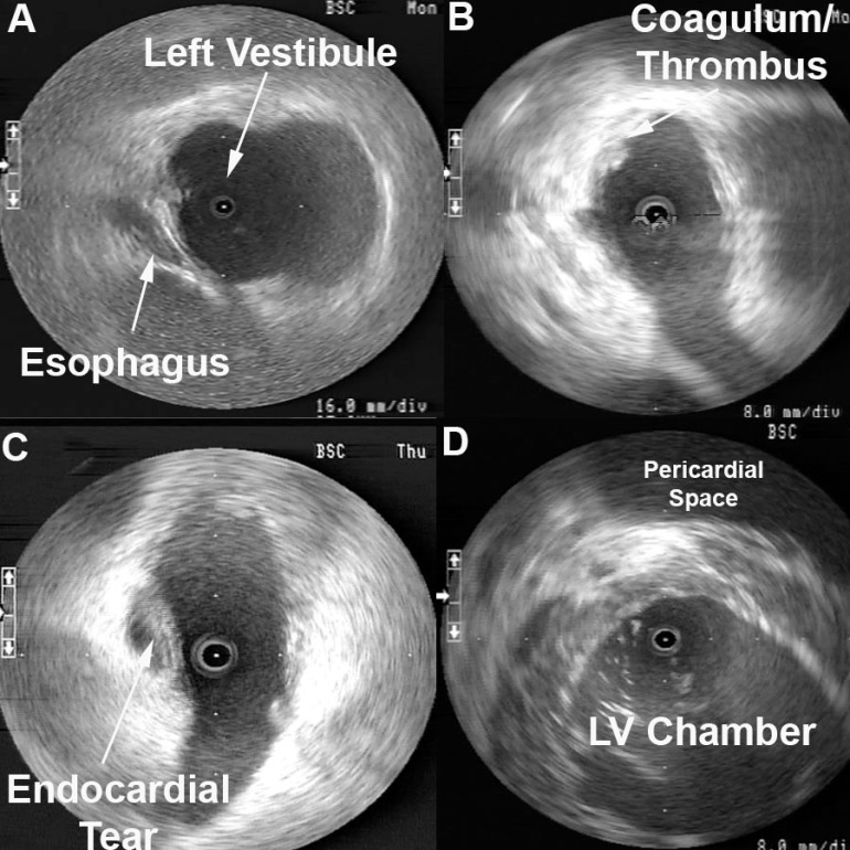
Esophageal Proximity can be monitored to guide locations of ablation to help minimize risk of esophageal damage. The entire length of esophagus that is contiguous with the left atrial posterior wall can be visualized with intra left atrial ICE to monitor ablation delivery and power titration. [1] Figure 1A shows the typical location of the esophagus during an atrial fibrillation ablation. Ablation over the esophagus is avoided or power is titrated to minimize risk of esophageal damage. Endocardial thrombi or coagulum can be detected using radial ICE as shown in Figure 1B. Left atrial damage can also be monitored using radial ICE. Figure 1C shows an unusual case of a tear or rent in the endocardium discovered during an atrial fibrillation ablation. Finally, though radial ICE is not the ideal imaging modality to evaluate for pericardial effusions given its limited far-field resolution. Figure 1D shows the pericardial space in view when an intra-left ventricular ICE position is utilized.

Figure 1 Radial ICE to Monitor for Intraprocedural Complications. Image A shows the left pulmonary vestibule with catheter evident at 9 o’clock and the esophagus viewed obliquely at ~7 o’clock. Image B shows a coagulum versus thrombus adherent to the endocardium. Image C shows a left atrial endocardial tear that did not result in pericardial effusion. Image D shows the pericardial space when ICE catheter positioned across the mitral annulus in the LV.
References:
1 Ren JF, Lin D, Marchlinski FE, Callans DJ, Patel V. Esophageal imaging and strategies for avoiding injury during left atrial ablation for atrial fibrillation. Heart Rhythm. 2006;3: 1156-1161.