Palpitations and Other Symptoms That May Represent Heart-Rhythm Abnormalities

This second podcast in the What are Palpitations? series focuses on palpitations and other symptoms that may represent heart-rhythm abnormalities. A brief outline includes:

  1. Introduction
  2. What is a normal heart rhythm?
  3. What are abnormal heart rhythms?
  4. What are palpitations?
  5. Other symptoms that may represent heart-rhythm abnormalities
  6. Know your ejection fraction
  7. What are ventricular fibrillation and ventricular tachycardia?
  8. Is there a difference between atrial fibrillation and ventricular fibrillation?
  9. Congestive heart failure and risk of arrhythmia

Please check back with the Heart Rhythm Center for future podcasts to include:

  • How Are Heart-Rhythm Abnormalities Diagnosed?
  • Work-up and Evaluation of Heart-Rhythm Disorders (Meeting the Heart-Rhythm Physician)
  • What Are the Common Supraventricular (Top-Chamber) Tachycardias?
  • What Is Atrial Fibrillation?
  • What Are the Common Ventricular (Bottom-Chamber) Tachycardias?
  • Treatment Options for Arrhythmias
  • The Electrophysiology Study and Ablation Procedure
  • Possible Complications of Electrophysiology Studies and Ablations
  • Postoperative Care after an EP Study (and possible ablation)
  • Psychosocial Impact of Arrhythmias

What are Palpitations? Introduction and Basics of Heart Anatomy


Thousands of patients every year are admitted to hospitals because of irregular, fast, or strong heartbeats. For the portion that will eventually be diagnosed with a heart condition, there are important choices to make moving forward. But there is rarely time in a short doctor’s visit to go over all the details.

The Heart Rhythm Center’s podcast What are Palpitations? serves as a comprehensive overview fills that information gap by imparting everything there is to know about abnormal heart rhythms through all stages of care. Dr. Jeffrey L. Williams, MD, MS, FACC, FHRS, CPE, has worked to provide patients with complete information. There are various types of arrhythmias, and the benefits and risks of treatments for each will vary for each individual patient. Patients, as well as their families, need a thorough understanding to make a fully informed decision.

Along with types of treatments, Dr. Williams discusses what is happening on a physiological level and explains the full evaluation process that doctors use. His knowledge, gained over years of study and practice, offers the what, why, and how of this medical issue, so anyone can make the best decisions for the health of his or her family.

Please check back with the Heart Rhythm Center for future podcasts to include:

  1. Palpitations and Other Symptoms That May Represent Heart-Rhythm Abnormalities (Arrhythmias)
  2. How Are Heart-Rhythm Abnormalities Diagnosed?
  3. Work-up and Evaluation of Heart-Rhythm Disorders (Meeting the Heart-Rhythm Physician)
  4. What Are the Common Supraventricular (Top-Chamber) Tachycardias?
  5. What Is Atrial Fibrillation?
  6. What Are the Common Ventricular (Bottom-Chamber) Tachycardias?
  7. Treatment Options for Arrhythmias
  8. The Electrophysiology Study and Ablation Procedure
  9. Possible Complications of Electrophysiology Studies and Ablations
  10. Postoperative Care after an EP Study (and possible ablation)
  11. Psychosocial Impact of Arrhythmias


Asystole Associated with Ablation near the Left Superior Pulmonary Vein

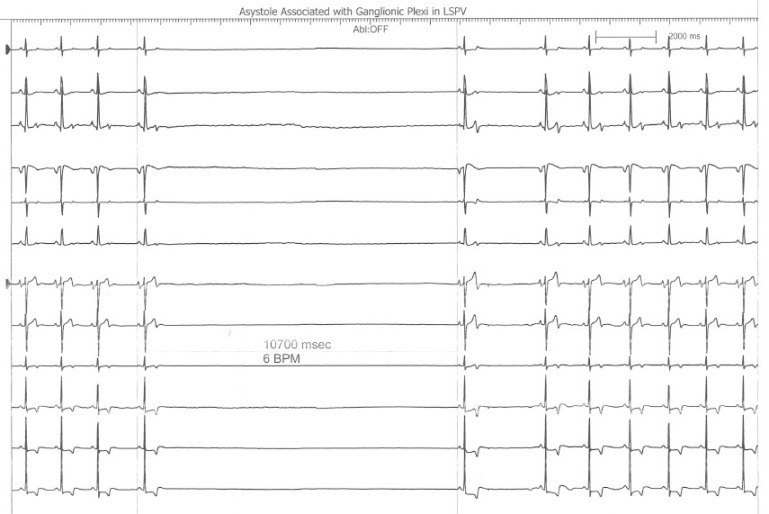
This is an interesting finding observed during a recent atrial fibrillation ablation performed in our Heart Rhythm Center.  The ablation paradigm has been previously described [1] and consists of a pulmonary venous antrum isolation using entrance and exit block criteria guided by intra left atrial radial intracardiac echocardiography (ICE). During the initial antrum encircling lesion asystole developed (see following figure), ablation was stopped, and sinus rhythm recovered within 10seconds.

Asystole During Ganglionic Plexi Ablation in LSPV

The following radial ICE image demonstrates the ablation catheter location in the superior aspect of the left pulmonary venous antrum near the left atrial appendage.

Radial ICE View LSPV Ganglion

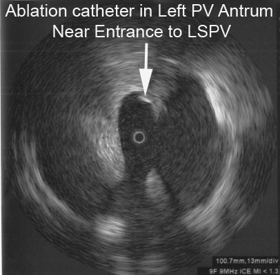
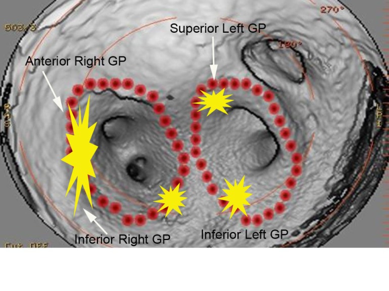
Bradycardia is often seen during atrial fibrillation ablations when proximate to autonomic ganglionic plexi.  [2]  I routinely see fluctuations in basal sinus rate during pulmonary venous antrum ablations but this was more dramatic than the sinus rate changes I usually observe.  This location as seen on the intra left atrial radial ICE shot is slightly more anterior than the left superior ganglionic plexus is usually expected.  The following figure shows a CT reconstruction of the posterior left atrium and pulmonary venous antra.  The red dots depict a typical venous antrum ablation lesion set and the yellow areas denote the approximate locations of the ganglionic plexi. [3]  Discontinuation of ablation led to quick restoration of sinus rhythm and repeat ablation near this location to finalize lesion set did not result in repeat asystole or significant fluctuations in sinus rate.


Approximate Locations of Ganglionic Plexi

Another possible explanation for this finding is acute sinus node dysfunction (from damage to the sinus node artery, SNA) during ablation in the anterior left atrium.  Chugh et al present an excellent review of coronary arterial injury during ablation of atrial fibrillation. [4]  Though there was no obvious PR prolongation prior to the pause suggesting an autonomic effect, there was also no obvious sinus tachycardia or acceleration serving as a “harbinger of impending [sinus node] dysfunction.”    Though the SNA arises from the RCA in two-thirds of patients, the remainder of SNA arise from an early branch of the circumflex which “passes superiorly and to the right of the LAA and courses over the anterior LA before terminating at the cavoatrial junction.”  Less commonly, the SNA branches off a more distal portion of the circumflex and ascends in the lateral ridge between the appendage and the left pulmonary veins.  The patient had an uneventful post-ablation recovery.


References:

1                     Schwartzman D, Williams JL, “On the Electroanatomic Properties of Pulmonary Vein Antral Regions Enclosed by Encircling Ablation Lesions,” Europace , V. 11 (2009), pp. 435–444.

2                     Pappone C, et al “Pulmonary vein denervation enhances long-term benefit after circumferential ablation for paroxysmal atrial fibrillation,” Circulation, V. 109 (2004), p. 327.

3                     Katritsis DG et al, “Autonomic Denervation Added to Pulmonary Vein Isolation for Paroxysmal Atrial Fibrillation A Randomized Clinical Trial,” JACC, V. 62 (December 2013), pp. 2318–25.

4                     Chugh A et al, “Manifestations of coronary arterial injury during catheter ablation of atrial fibrillation and related arrhythmias,” Heart Rhythm, V. 10, No. 11 (November 2013), pp. 1638-1645.

Three Dimensional Reconstruction of Left Atrium

The performance of complex cardiac procedures, such as advanced defibrillator placement, structural heart interventions, or arrhythmia ablation, is facilitated by the visualization of 3D anatomy.

     The performance of complex cardiac procedures, such as advanced defibrillator placement, structural heart interventions, or arrhythmia ablation, is facilitated by the visualization of 3D anatomy.  Providing 3D views of internal body structures and interventional devices in one image, this state-of-the-art system assists physicians in diagnosis, surgical planning, interventional procedures and treatment follow-up.     It permits better management of structural heart disease, streamlines interventional procedures, and minimizes radiation dose to physicians, staff and patients by selecting working views without fluoroscopy.  Patients can undergo 3D angiography of the coronary sinus to guide a biventricular defibrillator implantation with a left ventricular pacemaker lead.  Patients can also undergo 3D angiography of the left atrium and pulmonary veins to plan an atrial fibrillation arrhythmia ablation.