Free Registration for the Lakeland Regional Health Cardiovascular Symposium on February 25, 2017

We hope you will join us for Lakeland Regional Health’s 2017 Cardiovascular Symposium. We believe that you will find this opportunity to learn from leaders in our profession both educational and inspiring. Speakers from University of Pennsylvania, Vanderbilt University, University of Pittsburgh, and University of South Florida as well as local faculty will be presenting state-of-the-art topics in cardiovascular disease.

Lakeland Regional Health is committed to delivering nationally recognized healthcare, strengthening our community and advancing the future of healthcare. The experienced physicians of our Heart Center place patients at the heart of all they do. We are Polk County’s pioneer in expert cardiac care and have been for more than three decades.

Upon completion of our Symposium, participants should be able to:

  • Understand the latest research in managing patients with artificial hearts and/or ventricular assist devices.
  • Describe the long-term impact of cardiovascular care on function in the elderly.
  • Identify outpatients with pulmonary hypertension.
  • Recognize and describe the pros and cons of rate versus rhythm control for atrial brillation.
  • Identify and describe interventional cardiology technologies that are currently available to treat structural heart disease.
  • Describe the latest methods for outpatient management and diagnosis of peripheral vascular disease.
  • Describe the current inpatient and outpatient congestive heart failure care continuum.
  • Understand survival rates and long-term complications of adults with congenital heart disease.

    We look forward to seeing you in February. If you have any questions, please do not hesitate to contact us at 863.687.1190.

The Symposium offers 5.25 AMA Category 1 CME credits) and registration is free at 2017 Lakeland Regional Health Cardiovascular Symposium.

Asystole Associated with Ablation near the Left Superior Pulmonary Vein

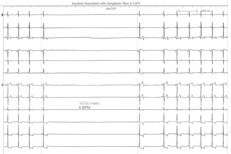
This is an interesting finding observed during a recent atrial fibrillation ablation performed in our Heart Rhythm Center.  The ablation paradigm has been previously described [1] and consists of a pulmonary venous antrum isolation using entrance and exit block criteria guided by intra left atrial radial intracardiac echocardiography (ICE). During the initial antrum encircling lesion asystole developed (see following figure), ablation was stopped, and sinus rhythm recovered within 10seconds.

Asystole During Ganglionic Plexi Ablation in LSPV

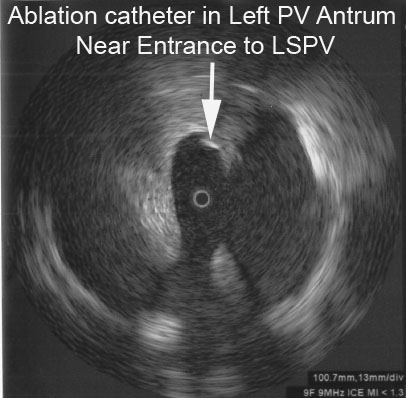
The following radial ICE image demonstrates the ablation catheter location in the superior aspect of the left pulmonary venous antrum near the left atrial appendage.

Radial ICE View LSPV Ganglion

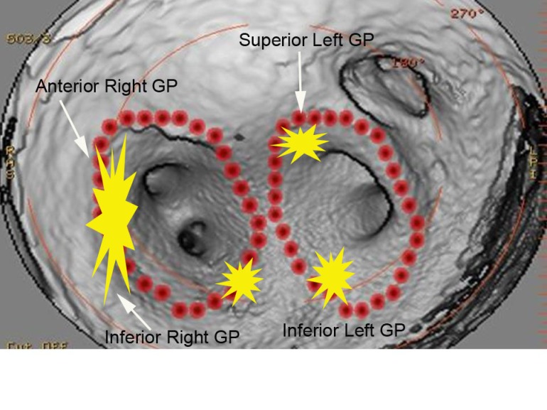
Bradycardia is often seen during atrial fibrillation ablations when proximate to autonomic ganglionic plexi.  [2]  I routinely see fluctuations in basal sinus rate during pulmonary venous antrum ablations but this was more dramatic than the sinus rate changes I usually observe.  This location as seen on the intra left atrial radial ICE shot is slightly more anterior than the left superior ganglionic plexus is usually expected.  The following figure shows a CT reconstruction of the posterior left atrium and pulmonary venous antra.  The red dots depict a typical venous antrum ablation lesion set and the yellow areas denote the approximate locations of the ganglionic plexi. [3]  Discontinuation of ablation led to quick restoration of sinus rhythm and repeat ablation near this location to finalize lesion set did not result in repeat asystole or significant fluctuations in sinus rate.


Approximate Locations of Ganglionic Plexi

Another possible explanation for this finding is acute sinus node dysfunction (from damage to the sinus node artery, SNA) during ablation in the anterior left atrium.  Chugh et al present an excellent review of coronary arterial injury during ablation of atrial fibrillation. [4]  Though there was no obvious PR prolongation prior to the pause suggesting an autonomic effect, there was also no obvious sinus tachycardia or acceleration serving as a “harbinger of impending [sinus node] dysfunction.”    Though the SNA arises from the RCA in two-thirds of patients, the remainder of SNA arise from an early branch of the circumflex which “passes superiorly and to the right of the LAA and courses over the anterior LA before terminating at the cavoatrial junction.”  Less commonly, the SNA branches off a more distal portion of the circumflex and ascends in the lateral ridge between the appendage and the left pulmonary veins.  The patient had an uneventful post-ablation recovery.


References:

1                     Schwartzman D, Williams JL, “On the Electroanatomic Properties of Pulmonary Vein Antral Regions Enclosed by Encircling Ablation Lesions,” Europace , V. 11 (2009), pp. 435–444.

2                     Pappone C, et al “Pulmonary vein denervation enhances long-term benefit after circumferential ablation for paroxysmal atrial fibrillation,” Circulation, V. 109 (2004), p. 327.

3                     Katritsis DG et al, “Autonomic Denervation Added to Pulmonary Vein Isolation for Paroxysmal Atrial Fibrillation A Randomized Clinical Trial,” JACC, V. 62 (December 2013), pp. 2318–25.

4                     Chugh A et al, “Manifestations of coronary arterial injury during catheter ablation of atrial fibrillation and related arrhythmias,” Heart Rhythm, V. 10, No. 11 (November 2013), pp. 1638-1645.

Detection of Atrial Fibrillation using iPhone 4S

Afib on Iphone Image taken from Heart Rhythm 2013;10:315-319.

A recent article in the Heart Rhythm Journal examines the performance of a smart-phone based application to detect atrial fibrillation. McManus et al conducted real-time analyses using two different statistical methods: root mean square of successive RR differences and Shannon entropy. They found that an algorithm combining both methods demonstrated excellent sensitivity (0.962), specificity (0.975), and accuracy (0.968) for beat-to-beat discrimination of an irregular pulse during afib from sinus rhythm. In an editorial, Dr. Dave Callans (from the University of Pennsylvania) congratulated the authors for this promising new strategy but raised questions on the clinical utility of this application in the absence of better strategies to manage and interpret this new data.

Ventricular Fibrillation after Elective Percutaneous Coronary Intervention

Case Description:

A 60 year old with past medical history of tobacco abuse was admitted for evaluation of chest pain without significant electrocardiogram (ECG) changes, electrolyte abnormalities or troponin elevation. Stress test revealed fixed inferolateral defects with EF44% and associated hypokinesis. Interestingly, an echocardiogram revealed an EF 55-60% with no regional wall motion abnormalities. Catheterization revealed an obstructive lesion in the PDA that had drug eluting stent successfully placed. Approximately one hour after stent placement routine ECG did not reveal any significant acute changes and patient was asymptomatic (see Figure 1). Approximately 150min after stent placement, the patient had an episode of ventricular fibrillation (VF) that required an external DC cardioversion (see Figure 2). Repeat cardiac catheterization did not reveal stent thrombosis or spasm. The patient underwent an uncomplicated single chamber defibrillator placement the following day.
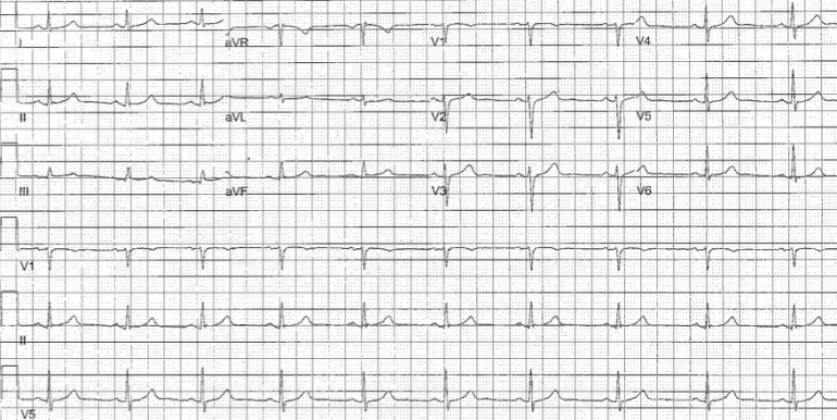
Figure 1.  EKG Prior to VF Arrest


Figure 2.  Telemetry Strip Showing the VF Arrest


Discussion:

VF arrest during PCI has been reported to have an incidence of 2.1% with higher incidence of VF during right coronary artery PCI. (HUA02) VF arrest during PCI is most commonly precipitated by contrast, ischemia from coronary dissection, embolism, spasm, or catheter manipulation and occurs during the cardiac catheterization. (NIS84) VF arrest after elective percutaneous coronary intervention (PCI) is uncommon. Indeed, an examination of 19,497 patients undergoing PCI revealed a 0.84% incidence of VF and no episodes of VF arrest temporally unrelated to vessel injection were reported. (ADD05) Survivors of VF arrest in the setting of myocardial infarction (MI) have similar mortality to those not experiencing VF arrest during acute MI. (DEJ09) In contrast, mortality in survivors of in-hospital cardiac arrest has been reported as high as 47% during a median followup of 1.3 years. (HEL11) It is unclear if the mechanism of VF arrest in our patient is secondary to PCI or rather a primary VF arrest. There is a prior report of delayed three vessel coronary spasm in a patient receiving paclitaxel drug-eluting stents however, coronary spasm was demonstrated on repeat catheterization in that report. (KIM05) Our patient did not report any ischemic symptoms preceding his VF arrest (though his EKG had subtle ST changes suggesting possible ischemia) nor did his repeat catheterization reveal vessel thrombosis, spasm, or dissection. Additionally, peri- and post-procedural myocardial injury from slow coronary flow, microvascular embolization, and elevated levels of troponin causing reperfusion tissue damage and cardiac dysfunction leads to worse long-term prognosis than those without myocardial injury (ISH08); our patient did not have significantly elevated pre- or post-procedure troponin levels. The time course of ischemia-induced reperfusion changes is likely less than 30minutes based upon prior experimental models of ischemia. (WIL08) Five minutes of coronary artery occlusion avoids increased risk of ventricular arrhythmias in animals and 30 minutes is appropriate for adequate reperfusion. (DAV81,DAV82,RUF79,WIL08) When the left anterior descending artery is transiently occluded in dogs, there is an initial (t=0-2minutes) small decrease in peak R wave amplitude and conduction velocity followed by a large increase in these indices over the ensuing 1-2minutes. (HOL76) There is a rapid return to baseline when occlusion is released and reperfusion occurs. This biphasic response has also been documented in dogs undergoing circumflex artery occlusions lasting 5minutes. (DAV81,DAV82) However, Ruffy et al (RUF79) found that LAD occlusions for 5 minutes in the dog resulted in a decrease in electrogram R wave amplitudes with no biphasic response. The progressive decrease in R wave amplitude (with the subsequent increase in amplitude) has been demonstrated in isolated rabbits hearts during global ischemia over 10 minutes (KAB89), isolated pig hearts during LAD occlusions for 5 minutesJAN86, and humans subject to 60minutes of unresolved ischemia. (VAI94) Of note, these electrical alterations were rapidly reversible upon reperfusion. (HOL76,RUF79,KAB89,JAN86) In summary then, our patient experienced a VF arrest 150min after elective PCI without conclusive evidence of procedural-related ischemia and well outside the conventional 30min window of reperfusion electrical alterations seen in experimental models. The role of defibrillator implantation as secondary prevention in patients like this is unclear.

References:

HUA02 Huang JL, Ting C-T, Chen Y-T, Chen S-A, “Mechanisms of ventricular fibrillation during coronary angioplasty: increased incidence for the small orifice caliber of the right coronary artery,” International Journal of Cardiology, Volume 82, Issue 3 (March 2002), pp. 221-228.

NIS84 Nishimura RA, Holmes DR Jr, McFarland TM, Smith HC, Bove AA, “Ventricular arrhythmias during coronary angiography in patients with angina pectoris or chest pain syndromes,” Am J Cardiol. V. 53, No. 11 (June 1984), pp. 1496-9.

ADD05 Addala S. Kahn JK, Moccia TF, Harjai K, Pellizon G, Ochoa A, O’Neill WW, “Outcome of Ventricular Fibrillation Developing During Percutaneous Coronary Interventions in 19,497 Patients Without Cardiogenic Shock,” Am J Card, V. 96 (2005), pp. 764-765.

HEL11 Helton TJ, Nadig V, Subramanya SD, Menon V, Ellis SG, Shishehbor MH, “Outcomes of cardiac catheterization and percutaneous coronary intervention for in-hospital VT/VF cardiac arrest, Catheter Cardiovasc Interv, [Epub ahead of print], (July 6, 2011).

DEJ09 DeJong JSSG, Marsman RFHenriques JPS, Koch KT, de Winter RJ, Tanck MWT, Wilde AAM, Dekker LRC, “Prognosis among survivors of primary ventricular fibrillation in the percutaneous coronary intervention era,” Am Heart J, V. 158, No. 3 (September 2009), pp. 467-472.

KIM05 Kim JW, Park CG, Seo HS, Oh DJ, “Delayed severe multivessel spasm and aborted sudden death after Taxus stent implantation,” Heart, V. 91, No. 2 (Feb 2005 Feb), e15.

ISH08 Ishiia H, Amanoa T, Matsubarabv T, Murohara T, “Pharmacological Prevention of Peri-, and Post-Procedural Myocardial Injury in Percutaneous Coronary Intervention,” Current Cardiology Reviews, V. 4. No. 3 (August 2008), pp. 223-230.

WIL08 Williams JL, Mendenhall GS, Saba S, “Effect of Ischemia on Implantable Defibrillator Intracardiac Shock Electrograms,” J Cardiovasc Electrophysiology, Vol. 19, No. 3 (March 2008), pp. 275-281.

DAV81 David D, Naito M, Chen CC, Michelson EL, Morganroth J, Schaffenburg M, “R-wave Amplitude Variations During Acute Experimental Myocardial Ischemia: An Inadequate Index for Changes in Intracardiac Volume,” Circulation, V. 63, No. 6 (June 1981), pp. 1364-1370.

DAV82 David D, Naito M, Michelson EL, Watanabe Y, Chen CC, Morganroth J, Schaffenburg M, Blenko T, “Intramyocardial Conduction: A Major Determinant of R-wave Amplitude During Acute Myocardial Ischemia,” Circulation, V. 65, No. 1 (January 1982), pp. 161-167.

RUF79 Ruffy R, Lovelace DE, Mueller TM, Knoebel SB, Zipes DP, “Relationship between Changes in Left Ventricular Bipolar Electrograms and Regional Myocardial Blood Flow during Acute Coronary Artery Occlusion in the Dog,” Circulation Research, V. 45, No. 6 (December 1979), pp. 764-770.

HOL76 Holland RP, Brooks H, “The QRS Complex during Myocardial Ischemia,” Journal Clinical Investigation, V. 57 (March 1976), pp. 541-550.

KAB89 Kabell G, “Ischemia-Induced Conduction Delay and Ventricular Arrhythmias: Comparative Electropharmacology of Bethanidine Sulfate and Bretylium Tosylate,” Journal Cardiovascular Pharmacology, V. 13, No. 3 (1989), pp. 471-482.

VAI94 Vaitkus PT, Miller JM, Buxton AE, Josephson ME, Laskey WK, “Ischemia-induced changes in human endocardial electrograms during percutaneous transluminal coronary angioplasty,” American Heart Journal, V. 127, No. 6 (June 1994), pp. 1481-1490.

JAN86 Janse MJ, “Electrophysiology and electrocardiology of acute myocardial ischemia,” Can J Cardiology, Supp. A (July 1986), pp. 46A-52A.
Floating Potential and Far-Field Left Atrial Appendage Signals During Atrial Fibrillation Ablation

Atrial fibrillation ablations are often like exploring a new forest; there are interesting findings unique to every patient and it is “what keeps you coming back.”  This is an electrogram recorded during an ablation after completing a left atrial pulmonary venous antrum isolation and assessing the left superior pulmonary vein for entrance/exit block. On first exam, it looks as if some work needs to be done but on closer inspection there is evidence of a “floating potential” and far-field sensing of atrial appendage activity.  The longer cycle length electrogram (~3100msec) is a pulmonary vein potential that is firing regularly but not conducting out to the left atrium (exit block).  In addition, the shorter cycle length (~1000msec) is a slightly lower frequency left atrial appendage signal that is often recorded from the proximate left superior pulmonary vein.  I get the “floating potentials” less than 10-20% of cases but it is a welcome finding.

ECG Sensing Driver’s Seat

Biomonitoring will become more and more common as we move forward.  The feasibility of us interacting with our electronic environment via biosensors will allow more personalized interactions.  We already see personalized interactions with:

1. Websites that track our preferences and suggest information that is related to these preferences.

2. Pacemakers and defibrillators that can adjust pacing rates based upon activity and breathing.

3. Glucose monitors that automatically adjust insulin dosing based upon blood glucose levels.

Ford is investigating the use of leadless ECG monitoring system in the driver’s seat.  Though it is investigational, one can see the possibility of tracking a driver’s heart for use in several ways such as monitoring for stress response prior to accident, ischemia, bradycardia (or heart block), and arrhythmias (such as atrial fibrillation).  Interesting to think about the implications of this data and privacy issues.

See link below for more information:

http://medgadget.com/2011/05/ford-unveils-contactless-ecg-sensing-driver-seat.html

Heart Rhythm Management and Drug Delivery via Implantable Biosensors

Your Next Prescription Might Be For A Microchip – Forbes.

Drug delivery via implantable biosensors is the next generation of devices that can be used to help heart rhythm disorders.  Our current implantable loop recorders allow us to track heart rhythm disorders and link symptoms to arrhythmias.  Implantable biosensors will combine this monitoring ability with automated drug delivery.

High Frequency Jet Ventilation During Ablation of Supraventricular and Ventricular Arrhythmias

High-frequency jet ventilation (HFJV) is used to decrease respiratory motion during atrial fibrillation ablations; however, the patient safety and efficacy of HFJV has not been evaluated during routine electrophysiology (EP) studies with radiofrequency ablation. This is a retrospective chart review of consecutive patients who underwent EP studies and ablations for supraventricular and ventricular arrhythmias while using HFJV. Any EP studies performed using HFJV where ablation was attempted were included for analysis; EP studies where no ablation was performed were not included. Patients underwent induction of general anesthesia with endotracheal intubation using intermittent positive pressure ventilation with sevoflurane in the EP laboratory prior to vascular access. HFJV was then provided by a commercial system with initial settings: ventilation rate at 100 cycles/min and driving pressure at 20–25 psi. Total intravenous anesthesia was then provided with dexmedetomidine and propofol as well as fentanyl and rocuronium titrated to bispectral index (Bis) score <60. The overall mean age of patients (n=72) was 55+/-18 years (ranges 18–84 years). The mean creatinine (mg/dl) was 1.0+/-0.3, the mean ejection was 0.58+/-0.08, and mean post-EP study length of stay was 1.4+/-0.9 days (range 1–5 days). There were no intraprocedural or major complications. There was a 6.9% rate of minor complications (n=5). There was a 97.2% overall ablation success rate (70 of 72 ablations). Ablations were successful in all subjects except for one left atrial flutter and one right atrial tachycardia. Only one of 72 (1.4%) procedures required discontinuation of general anesthesia and HFJV to induce arrhythmia (right ventricular outflow tract ventricular tachycardia). No patient experienced procedural awareness and the mean Bis score was 40+/-5.3. This report provides further evidence the routine use of jet ventilation in the electrophysiology laboratory is safe, well tolerated, and efficacious, with ablation success rates similar to traditional sedation/ventilation techniques with a variety of arrhythmias.

For Full Study Please See:  http://www.innovationsincrm.com/cardiac-rhythm-management/2011/november/157-high-frequency-jet-ventilation

Implantable Biosensors and Future Role in Electrophysiology

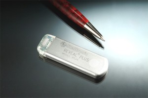
Implantable loop recorders (ILR) are used for long-term arrhythmia monitoring inpatients that have had syncope or cryptogenic strokes (possible from atrial fibrillation).  There are two primary models of loop recorders: St. Jude Medical’s Confirm (http://www.sjmprofessional.com/Products/US/Implantable-Cardiac-Diagnostics/SJM-Confirm-Implantable-Cardiac-Monitor.aspx) and Medtronic’s Reveal (http://www.medtronic.com/for-healthcare-professionals/products-therapies/cardiac-rhythm/cardiac-monitors-insert/reveal-dx-and-reveal-xt-insertable-cardiac-monitors-icms/index.htm).

ILR are placed just under the skin in the left chest and are able to record a patient’s heart rhythm.  It helps to diagnose the cause of syncope (fainting) or any number of heart rhythm disorders.  Initially, doctors try to diagnose heart rhythm disorders with monitors that are worn for 48hours to 2-4weeks; however, an arrhythmia that may only occur every few months will not be detected by a brief snapshot in a patient’s life.  ILR generally last 2-3 years before the battery wears out.

The exciting future of implantable monitors is not simply heart rhythm diagnosis.  Recently, the company MicroCHIPS announced results of the first human clinical trial of an implantable, wireless microchip drug delivery device (http://www.mchips.com/technology.html).  This type of implantable device can allow automated drug delivery over a period of several years.  One can imagine a device that can track a person’s heart rate and dose medication to permit precise control of heart during an abnormal heart rhythm.